An Algorithm from Netflix Challenge to Speed Up Biological Imaging?
Raman spectroscopy is a well-known non-invasive technique for determining the chemical composition of complex samples. For example, it has shown promise for the identification of cancer cells, as well as the analysis of tissues in search of pathologies. Although this method is remarkably simple (no sample preparation is required), capturing the rapid dynamics of biological samples requires an unfeasible acquisition rate. In addition, processing the huge amount of data generated by spectroscopic imaging is time-consuming, and often a limitation to studying specimens over large areas.
As part of an international collaboration involving LPENS and LKB, a methodology has been developed to simultaneously address these two challenges: increasing the sampling rate and introducing a simpler way to obtain useful information from spectroscopic images. This work was published in the open-source review Optica.
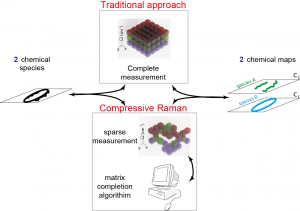
To speed up the process, the researchers modified their Raman system by replacing the slow and expensive cameras with a fast and inexpensive spatial light modulator based on micromirror arrays. This device allows to select wavelength groups, which are then picked up by a highly sensitive single pixel detector.
This device deliberately acquires only a fraction of the data required for conventional Raman spectroscopic imaging, compressing the images as the acquisition progresses. Then, the missing data are completed by using a very special calculation: an adaptation of the algorithm originally developed for predicting Netflix’s (2009) cinematographic preferences!
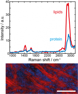
The researchers tested their new imaging methodology on brain tissue and individual cells, both of which are chemically highly complex. The results showed that this technique allows images to be acquired in a few tens of seconds, whereas the traditional Raman approach generally took a few minutes. At the same time, they compress the acquired data, which reduces their volume by a factor of 64!
The researchers believe that this new approach should work with most biological samples, but they plan to test it with other types of tissues to generalize their demonstration. In addition to clinical tools, the method could be useful for many biological applications, such as seaweed characterization. Similarly, they also want to improve the scanning speed of their system to achieve an acquisition speed of less than one second. Such a breakthrough would allow the use of the algorithm for clinical applications such as tumor detection or tissue analysis.
Hilton B. de Aguiar, coordinator of the collaboration, holds a Junior Research Chair in the Physics Department of the ENS.

Caption: Compressive Raman Imaging – With the new compressive Raman approach, one can acquire less spectral data than traditionally required and then use the matrix completion algorithm to fill in information not recorded.

