
IMAGING IN COMPLEX MEDIA
from wavefront shaping to computational imagingPublication(s)
- O.Katz, P.Heidmann, M.Fink, S. Gigan, Non-invasive real-time imaging through scattering layers and around corners via speckle correlations, Nature Photonics, 8, 794 (2014) link
-
T. Wu, J. Dong, X. Shao, S. Gigan Imaging through a thin scattering layer and jointly retrieving the point-spread-function using phase-diversity Opt. Express 25, 27182 (2017) link
-
T. Wu, O. Katz, X. Shao, S. Gigan Single-shot diffraction-limited imaging through scattering layers via bispectrum analysis Opt. Lett. 41(21), 5003 (2016) link
imaging behind scattering layers
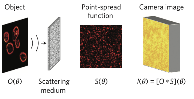 For a surface scatterer or a thin scattering system, there is an angular “memory effect” of the speckle. Consequently, all information is not completely lost when the image of an object propagates through such a medium. Using a technique known as “phase-retrieval”, we showed in 2014 that, owing to the ‘memory-effect’ for speckle correlations, a single high-resolution image of the scattered light, captured with a standard camera, encodes sufficient information to image through visually opaque layers and ar
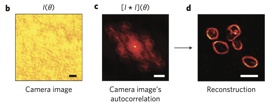 ound corners with diffraction-limited resolution. we show that the autocorrelation of the scattered light pattern is essentially identical to the autocorrelation of the object’s image itself. We then reconstruct the object’s image from its autocorrelation using an iterative Fienup-type algorithm . As proofs of concept, we experimentally demonstrate our single-shot non-invasive technique through a variety of highly scat- tering samples, from optical diffusers to dynamically varying biological tissues. In addition, we demonstrate imaging ‘around corners’ by recording the diffuse light back-scattered off white- painted ‘walls’.
Our more recent work focus on making the technique more robust and more adapted to realistic scenarios.
Wavefront Shaping in biological tissues
(collab. L. Bourdieu @IBENS)
we explore the possibility of improving image quality for deep microscopy in biological tissues, which are typically both forward scattering and aberrating. Unfortunately, conventional wavefront sensing techniques are not usable anymore. Until 2014, we used a both conventional adaptive optics setup, with a traditional deformable mirror, and optimize deep imaging using the image quality itself as a metric, trying to improve directly conventional deep imaging techniques such as Optical Coherence Tomography.
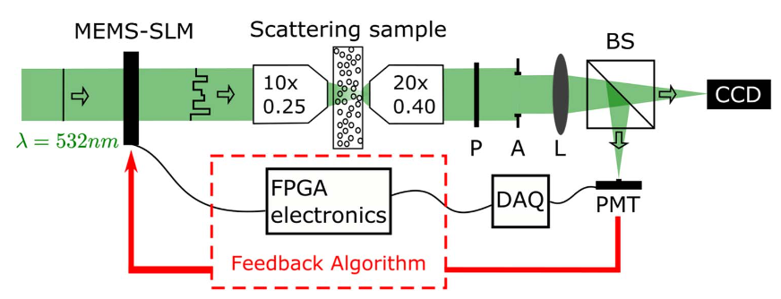 More recently, we started exploring the possibility to directly exploit wavefront shaping. For instance, we recently demonstrated the fastest (at the time) continuous optimization wavefront shaping system, able to focus light through dynamic scattering media. A micro-electro-mechanical system-based spatial light modulator (from Boston Micromachine Corp), a fast photodetector,
 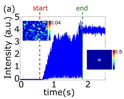 and field programmable gate array electronics were combined to implement a continuous optimization of a wavefront with a single-mode optimization rate of 4.1 kHz.
The system performances were demonstrated by focusing light through colloidal solutions of TiO2 particles in glycerol with tunable temporal stability.
An important aspect of biological tissues is their forward scattering properties. We in particular studied how the memory effect, a spatial correlation very useful for imaging, was actually much larger than expected in biological tissues.
Publications
- Blochet, K. Joaquina, L. Blum, L. Bourdieu, S. Gigan Enhanced stability of the focus obtained by wavefront optimization in dynamical scattering media arXiv:1905.03757 [physics.optics]
- B. Blochet, L. Bourdieu, S. Gigan Focusing light through dynamical samples using fast closed-loop wavefront optimization Opt. Lett. 42, 4994 (2017) link
- S.Schott, J.Bertolotti, J.F. Léger, L. Bourdieu, S. Gigan Characterization of the angular memory effect of scattered light in biological tissues Optics Express 23,13505 (2015) link
- J. Binding, J. Ben Arous, J.F. Léger, S. Gigan, A.C. Boccara, L. Bourdieu, In vivo rat brain refractive index measurement using full-field OCT and consequences for two photon microscopy, Optics Express 19, 4833 (2011) OE
Publication(s)
A. Liutkus, D. Martina, S. Popoff, G. Chardon, O. Katz, G. Lerosey, S. Gigan, L. Daudet, I. Carron, Imaging With Nature: A Universal Analog Compressive Imager Using a Multiply Scattering Medium, Sci. Rep. 4, 5552; DOI:10.1038/srep05552 (2014).
Computational imaging : Exploiting disordered media for compressive sensing
(Collab. L. Daudet@ , F. Krzakala@ LPENS)
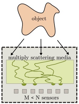
Compressive sensing (CS) is a paradigm change in signal acquisition, allowing to beat the Shannon-Nyquist sampling rate limit. If an image is sparse (i.e. compressible, which is the case for most natural objects) then CS shows that it can be sampled and retrieved in much less measurements than the Shannon-Nyquist theorem. It however requires that all measurements contain information about the whole object. Random media, as optimal mixers for light, are good candidate to perform such tasks. We showed that a calibrated random medium (calibrated by its transmission matrix) can be seen as an universal compressive sensor, and demonstrate compressive imaging.
Publications
- T. Chaigne, O. Katz, A.C. Boccara, M. Fink, E. Bossy, S. Gigan, Controlling light in scattering media noninvasively using the photo-acoustic transmission-matrix, Nature Photonics, 8, 58–64 (2014) NATPHOT ARXIV
- T. Chaigne, J. Gateau, O. Katz, E. Bossy, S. Gigan, Light Focusing and Two-Dimensional Imaging Through Scattering Media using the Photoacoustic Transmission-Matrix with an Ultrasound Array, Optics Letters, Vol. 39, Issue 9, pp. 2664-2667 (2014)
Photoacoustic focusing and imaging inside a scattering medium (collab. E. Bossy)
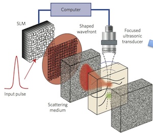 The ability to use wavefront shaping techniques for deep biological imaging, cannot rely on access to both side of a medium. One wants to work from one side only, and cannot place a detector inside the medium. Guiding light inside a complex medium by wavefront shaping requires a “guide star” or “beacon” to measure light intensity in depth. Acoustics wave, that propagates almost undisturbed in biological tissues are good candidates to probe light intensity. In our approach, we rely on photoacoustics, a technique where local light absorption deep inside a tissues generates an acoustic signal that can be detected and localized from the outside using a transducer. We demonstrate the measurement of a photoacoustic transmission matrix, that allow focusing in depth.